Introduction
Acute iodide sialadenitis, or “iodide mumps,” is an adverse
reaction to iodinated contrast media causing salivary gland swelling minutes to days after iodine exposure [1]. The prognosis of
iodide mumps is benign. Clinical features persist for 4-hours to
14-days, with complete healing without sequelae [2].
Several studies have suggested that this condition may be
underdiagnosed, with an incidence of 1-2% [3] and less than 80
cases reported in the literature to date [4]. Originally thought to
be IgE-mediated, the swelling symptoms have been mistakenly
attributed to angioedema or anaphylaxis [3]. The condition is
thought to represent a response to iodine accumulation in the
salivary glands, however the exact pathogenesis is still unclear [2].
Lack of physicians’ familiarity with this condition might result
in underdiagnosis, excessive diagnostic workup and inappropriate
treatment which therefore implies a misuse of resources and the
risk of additional iatrogenesis [3].
Case report
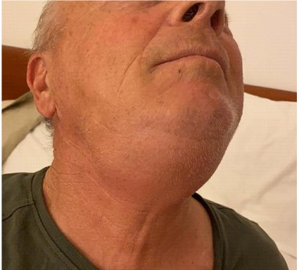
A 60-year-old patient with stage IIIA lung adenocarcinoma performed a Computed Tomography (CT) scan after completion of 4
courses of induction chemotherapy. After dinner, 12-hours after
contrast media injection, he reported swelling of the submandibular salivary glands. Physical examination revealed bilateral
submandibular salivary glands swelling, with mild tenderness at
palpation (Figure 1). Submandibular salivary glands swelling disappeared 24-hours after contrast media injection.
At the next follow up CT scan, 4 months later, the patient experienced the same symptoms, which disappeared with no treatment (Figure not shown).
At the further follow up CT scan, 10 months later, the patient
was advised to receive premedication with methylprednisolone
32 mg 12 hours and 2 hours before CT scan. With this premedication, the patient did not experience iodide mumps after iodinated
contrast media injection.
Discussion
Iodide mumps is a rare adverse reaction to iodine-containing
contrast media administration, characterized by a rapid, usually
painless enlargement of salivary glands [2].
A meta-analysis on the published case reports of iodide mumps
in the medical literature identified 77 cases of iodide-induced sialadenitis. Median age was 63 years, and 61% (47/77) were males.
Median time to onset was 16 hours, and symptoms resolved in a
median of 3 days. Twenty-seven subjects (35%, 27/77) were reported to have an impaired renal function at baseline. Symptoms
were resolved in all cases over a median of 3 days with no statistically significant difference between those who received therapeutic intervention and those who did not (p = 0.430). Older age
and longer time to onset were significantly associated with longer
duration of symptoms, and presence of tenderness demonstrated
statistically significant association with longer duration of symptoms [4].
Lucarelli et al conducted a review of the medical literature and
found out approximately 50 cases of iodide mumps. Of these, 29
cases involved the submandibular glands, and 15 cases involved
also the parotid glands. This can be supported by the observation
that the submandibular glands have a more viscous, prominently
mucin-rich, secretion [2]. Saro-Buendía et al conducted a prospective observational study. During the 2-months study period,
4 cases of contrast-induced sialadenitis were detected. Patients
were aged 68-76 years and presented a bilateral submandibular
gland swelling debuting 12 to 72 hours after an exposure to iodinated contrast. Characteristic ultrasonographic findings supported the diagnosis and the clinical course was self-limited after 60
to 150 hours [5].
The rarity of iodide mumps may hinder prompt diagnosis and
appropriate treatment.
Differential diagnosis is with an overt allergic reaction, which
can be ruled out by the lack of specific symptoms such as rash,
angioedema, dyspnea and hypotension. Furthermore, anaphylactic reactions usually occur within 30 minutes after exposure
to the antigen [2]. A wide variety of methods have been used to
treat iodide sialadenitis, including antihistamines, corticosteroids,
hyperhydration, and dialysis in patients with renal failure, none
of which have proven efficacy [3]. Current treatment for iodide
mumps consists in supportive therapy and administration of anti-inflammatory agents, whereas the role of steroids is still controversial [2]. In a case report, a 59-year-old white female with a diagnosis of iodine-related sialadenitis was given 20 mg of decadron
intravenously, with prompt resolution of the swelling within a few
hours [6]. In our patient, premedication with methylprednisolone
32 mg 12 hours and 2 hours before CT scan proved effective to
avoid recurrence of iodide mumps after CT contrast media injection.
The prognosis of iodide mumps is benign. There have been no
reported life-threatening complications of the condition to date
[2].
Conclusion
In conclusion, iodide mumps is a rare but benign adverse reaction to iodinated contrast media injection. Because of the widespread use of iodinated contrast-enhanced imaging, such as CT,
and interventional techniques, clinicians should be aware of this
entity to avoid more aggressive diagnostic workup.
Conflicts of interest: The authors declare that they have no
conflicts of interest.
References
- Afshar M, Alhussein M. Iodide-associated sialadenitis. N Engl J
Med. 2017; 376: 868.
- Lucarelli A, Perandini S, Borsato A, Strazimiri E, Montemezzi S. Iodinated contrast-induced sialadenitis: a review of the literature and
sonographic findings in a clinical case. J Ultrason. 2018; 18: 359-364.
- Egan M, Maglione PJ. Multiple reasonably tolerated percutaneous
coronary interventions in a patient with iodide mumps. Ann Allergy Asthma Immunol. 2015; 115: 253-254.
- Jiao A, Farsad K, McVinnie DW, Jahangiri Y, Morrison JJ. Characterization of Iodide-induced Sialadenitis: Meta-analysis of the Published Case Reports in the Medical Literature. Acad Radiol. 2020; 27: 428435.
- Saro-Buendía M, Torres-García L, Mossi Martínez C, Battig Arriagada E, Carreres Polo J, et al. Management of iodine contrast induced salivary gland swelling (sialadenitis): experiences from an
observational study. Acta Otolaryngol. 2023; 143: 64-69.
- Alkaied H, Harris K, Azab B. A complete resolution of sialadenitis induced by iodine containing contrast with intravenous dexamethasone infusion. Clin Med Insights Gastroenterol 2012; 5: 61-63.