Introduction
Human Hepatocellular Carcinoma (HCC) is the primary cancer of the liver, which is the third-highest form of cancer and one of the leading causes of cancer death worldwide. It originates from latent chronic liver disease and cirrhosis. Cirrhosis is a chronic inflammation of the liver which leads to the formation of small nodular clusters of abnormal cells. An ideal environment for malignant, or cancerous tumors to grow is an inflamed nodular liver. Presently, there is a very limited scope of therapeutic measures for the advanced HCC treatment, and its response to any kind of chemotherapy is very poor, primarily due to the high level of chemically acquired and intrinsic immuno-resistance. Hence, the research to find out innovative and effective medical approaches for the treatment of such patients is very significant. A key ingredient, Berberine present in Hydrastis canadensis, has shown a critical anti-cancer effect in vivo as well as in vitro (Yu et al. 2014). Many studies have already shown that immortalized hepatic cell lines can be utilized for research and academic purposes instead of biopsy tissues of the liver, because of the high maintenance charge with the requirement of expertise handling, high sensitivity, decreased action of several key enzymes, and preserving of liver biopsies in an appropriate culture is challenging for research related works [1].
Goldenseal (Hydrastis canadensis L.) is a perennial, herbaceous plant of the Ranunculaceae family. In addition, the different plant parts, especially the rhizome contains numerous alkaloids namely, berberine, palmatine, hydrastine, hydrastinine, and canadine [2], and lesser amount of flavonoids (e.g. sideroxylin, 8-desmethyl-sideroxylin, 6-desmethyl-sideroxylin, etc), organic acids (e.g. neochlorogenic acid, chlorogenic acid, etc). The increasing demand for non-toxic phytochemical focusing therapies in the modern world has been the driver for research-related activities for promoting public health concerning issues [3]. The presence of active key constituents in ethanolic extract of alternative medicines has been a challenging area in the medical as well as the research community. In this study, we have used commercially available diluted homeopathic remedies for immortalized hepatic cell line HepG2 [4].
Apoptosis is a specific form of cell suicide or Programmed Cell Death (PCD), and among several markers of apoptosis, DNA fragmentation is considered a hallmark. Defect in apoptotic pathways plays a crucial part in the progression and development of cancer. Modulation of the apoptotic signaling pathways employing alternative treatment strategies can knock out cancer cells [5].
In general, cancer is a metabolic disorder that is primarily denoted by inflammation of body parts. Inflammation itself is a complicated procedure resulting from the secretion of numerous plasma and cellular mediators, like cytokines, which get released in response to an inflammatory stimulus or any injury [6].
Cytokines are cell-derived protein molecules that mediate immune and inflammatory reactions. There are two kinds of cytokines, anti-inflammatory, and pro-inflammatory cytokines. Chronic inflammation is caused by the secretion of pro-inflammatory mediators [7], and it has been studied that chronic low-grade inflammation is directly related to immunological disorders [8]. The inflammatory response is dictated by the balance between anti- and pro-inflammatory mediators. The pro-inflammatory cytokines like IFN ɣ, IL-6, IL-8, IL-1 β, and TNF α are authoritative for early and increased inflammatory reactions, and on the other hand, the anti-inflammatory cytokines, which include IL-10, TGF β1, and TGF β3, have the opposite effect. It has already been studied in vivo as well as in vitro that growth factors and cytokines are responsible for regulated gene expression and are mainly expressed by non-parenchymal cells like endothelial, transforming hepatic stellate cells, and Kupffer cells. But more clinical research is required about their regulation and production in parenchymal cells, and many parenchymal cell functions are retained by the human hepatocellular carcinoma cell line HepG2 [9]. In this study, we have tried to investigate the mortality rate and the gene exexpression patterns in a human HCC cell line (HepG2) in response to treatment with an extremely low concentration of the Goldenseal extract.
Materials and methods
Cell culture
The human hepatocellular carcinoma cell line HepG2 was procured from the cell repository of the National Centre for cell science, S.P. Pune University Campus. All cells used in this work were between passages 25 and 27. The HepG2 cell line was cultured in a T25 flask. The culture medium used was, Dulbecco’s modified Eagle’s medium (DMEM) (Gibco; Catalogue No. 10-566-016), along with 10% fetal bovine serum (FBS) (Gibco; Catalogue No. Catalogue No. 11573397), 10% F12 supplement (Gibco, and 400 μL antibiotic (Pen-Strep +Amphotericin) (Gibco, Lot No. 2321103), and cultured at 37°C in a humidified condition containing 5% CO2. Media was changed every 48 hrs to facilitate better growth. Cells were maintained at 37°C, at 5% CO2 atmosphere, and passaged before reaching confluence.
Source of the drug
The 6C dilution of Hydrastis canadensis drug was procured from Hahnemann Publishing Company Private Limited (HAPCO), Kolkata, a Government recognized manufacturing unit [Hydrastis Canadensis 6, HAPCO, M.L.HL 8M, Batch no. 0107, Manufactured in January 2022. (E-mail: hapco@vsnl.net)].
For replicating and further investigation of the research work, the 6C dilution of the drug can be prepared in-house following the below-mentioned procedure. The extract was imported by HAPCO (India) from Europe. The plant is the same as the herbarium accession number 154997, in Butler University Friesner Herbarium, collected by P. A Scott and the collection number is 2515. The native range of the plant is in Eastern North America. Preferably, the root (rhizome) portion of the plant is used for making the homeopathic mother tincture. The procedure for homeopathic potentization involves the dilution of the mother tincture (crude ethanolic extract of the plant material) in 99 mL of rectified spirit (90% ethyl alcohol) and given 10 succussions to produce the potency 1C. Similarly, 1 mL of the drug solution at potency 1C is again added with 99 mL of 90% ethanol, and 10 succussions given to produce the potency 2C in this way by successive dilutions and successions, further potencies like 6C and beyond are produced [10].
Cell viability assay
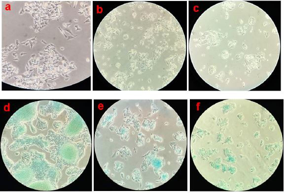
After being treated in three experimental sets, that is, i) Control (untreated cells), ii) Succused 70% Alcohol treated cells, and iii) Hydrastis canadensis 6C treated cells, for 24 hrs, the cells were photographed and used for methylene blue staining assay and MTT assay separately to detect morphological changes and proliferation rate respectively.
Methylene blue staining assay
The HepG2 cells were taken from the growth culture medium and diluted in a fresh culture medium. From this dilution, 3×10⁵ cells/mL was seeded into each respective well of a 6-well tissue culture plate. After 24 hrs of incubation at a condition of 37°C in 5% CO2, the media was discarded, followed by the addition of 1 mL methylene blue solution (1X PBS[Phosphate Buffer Saline] + 0.6% methylene blue + 1.25% glutaraldehyde) to each well, for cell staining and fixation. After 1 hr of incubation at 37°C in 5% CO2, the stain was pipetted out from each well and carefully rinsed with 1x PBS, and then images were captured (Figure 1). The methylene blue staining assay offers the advantage of fixing and staining the viable cells in the wells where they were cultured, while the dead cells are removed with the PBS wash before capturing the images [11].
MTT colorimetric assay
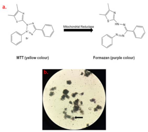
Cell viability assay was carried out using 3-(4,5-dimethylthiazol-2-yl)-2,5-diphenyltetrazolium bromide (MTT) [12],with minor modifications. The principle of this assay is that the tetrazolium salt (MTT) gets reduced to insoluble formazan crystals (Figure 2) by mitochondrial reductase enzyme activity in the live cells, whereas the dead cells cannot perform this activity. So, live cells with active metabolism can convert the MTT reagent into a deep purple-colored formazan product (Figure 1b). Hence, the intensity with which the colored product is formed is directly proportional to the count of live cells present in the culture medium [13,14]. The HepG2 cells were diluted in a fresh culture medium, and from this dilution 3×10⁵ cells/mL were seeded into each well of a 96-well plate and then incubated for 48 hrs in a 5% CO2 incubator at 37∘C. After 48 hrs, the alcohol and Hydrastis canadensis 6C medicine were added at 1: 10 dilution into the wells and then incubated for 1 hr. After 1 hr, 10μL of MTT reagent (EZcountTM, HiMedia) was added, and the mixture was further incubated for 4 hrs. Next, the mixture in each well was removed, and formazan crystals formed were dissolved in 100 μL of solubilization solution as provided in the kit and left for overnight incubation. After that, spectrophotometric measurement of the mixture was performed in Readwell Touch ELISA Plate Reader at wavelengths of 546 and 600 nm.
DNA ladder assay
The DNA ladder (DNA fragmentation) assay for apoptosis, following treatment with the Hydrastis canadensis 6C drug on the HepG2 cell line, was assessed by agarose gel electrophoresis [15,16]. Briefly, HepG2 (>90% confluency; 3×10⁵) cells were exposed to Hydrastis canadensis 6C drug for 24 hrs. Alcohol-treated and untreated control samples were also included in the experiment. After 24 hrs of incubation in a 5% CO2 incubator at 37°C, the used-up growth medium was discarded, and 1x PBS wash was done to get rid of the dead cells. Then 1 mL of trypsin was added to the cells and incubated for 10 mins in a 5% CO2 incubator at 37°C. After that 1mL of the growth medium was added to stop the trypsin activity. Cells were collected using a cell scraper and centrifuged at 1200 rpm for 10 min. The cell pellet was washed twice with 1x PBS and resuspended in 20 μL proteinase K and incubated for 1 hr at 56°C water bath. Then DNA was isolated by the salting-out method and quantified by a spectrophotometer, NanoDrop 2000 (Manufacturer: Thermo Scientific™ ND2000USCAN). The DNA samples were electrophoresed on a 1% agarose gel containing 4 μL/100 mL SYBR-Safe DNA gel stain (Invitrogen, Cat No. S33102). A 100 base pair (bp) DNA ladder (Trackit, Invitrogen, Cat No. 10488058) was used in the assay. The gel was examined and photographed by an ultraviolet gel documentation system (ChemiDoc™ XRS+ System with Image Lab™ Software; 1708265).
Analysis of gene expression
RNA extraction was performed from the HepG2 cells, using RNA isoPus reagent (Takara, Japan) following the manufacturer’s instructions. The RNA quality check was done by measuring the absorbance ratio at 260/280 nm of the samples. Purified RNA was reverse transcribed immediately after extraction, using the 5x iScript RT cDNA supermix (Biorad), and a quantity of 1 μg total RNA was added into a 20 μL reaction volume to synthesize cDNA. Quantitative Real-Time PCR was done, using the SYBR Green method (iTaqTM Universal SYBR Green Supermix, Biorad). The Housekeeping B-actin gene was used as an endogenous control. The following pre-designed primers were used as shown in (Table 1).
RT-PCR was done using the real-time SYBR green method following the cycling conditions as mentioned in (Table 2), and then the level of relative expression of mRNA was determined using B-actin as a housekeeping gene following the comparative 2−ΔΔCTcalculationsformula [17].
Test Experiment-Housekeeping experiment=ΔCT1;
Test control-House-keeping control=ΔCT2;
ΔCT1-ΔCT2=ΔΔCT; Gene Expression=2^-(ΔΔCT).
And the mean data of two replicates is represented using R-programming language (RStudio-2021.09.1.0).
Statistical analysis
Statistical analysis was executed using one-way Analysis Of Variance (ANOVA) followed by Tukey’s multiple comparison post-test, done using GraphPad Prism version 9.3.1 Software, for representation of MTT colorimetric assay for which the data is represented as Mean ± Standard Deviation (SD). The data were checked for normality and outlier removal and residuals before comparing the results.
Results
Methylene blue staining assay
The cell viability of HepG2 cell culture treated with Hydrastis canadensis 6C and alcohol was checked by methylene blue assay microscopically under 20x (TCM 400), and it was observed that stained live cells were maximum in control wells, less in alcohol-treated wells and least in drug-treated wells (Figure 1), the dead cells were removed before capturing images by 1x PBS wash.
MTT colorimetric assay
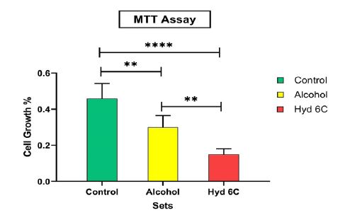
The cell viability (%) of HepG2 cells in response to Hydrastis Canadensis 6C and alcohol is presented as a bar graph in (Figures 2-4). The plot shows that there is a decrease in cell viability in the cell culture treated with the drug (P<0.0001), while cell viability was maximum in the control where the cells were not treated with any drug or alcohol. There was moderately decreased cell viability (P=0.0047) observed in the cell culture treated with alcohol, compared to the control set. It was observed that the cell viability remains intact in control but in alcohol and particularly in drug-treated cultures, the cell viability decreases, and a significant (P=0.0075) difference lies between the drug and alcohol. Thus it was observed that 6C dilution of Hydrastis canadensis effectively kills the cancer cells much more as compared to only alcohol treatment, which confirms the anti-cancer activity of the drug Hydrastis canadensis 6C.
DNA ladder assay
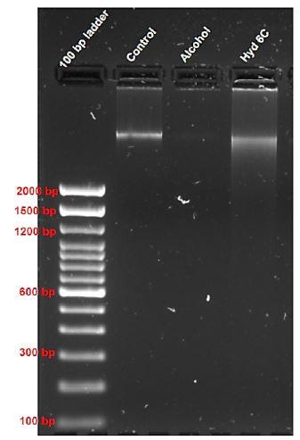
The alternative medicine-Hydrastis canadensis 6C showed significant cytotoxic activity on the HepG2 cells, so these were further tested for their ability to induce DNA fragmentation in-vitro using a DNA ladder assay. As shown in Figure 4, the band observed for the extracted DNA from the control set was intact, while a very faint band was observed for the extracted DNA from the alcohol-treated cells. On the other hand, a smear band pattern was observed for the extracted from the Hydrastis canadensis 6C treated cells. Thus DNA fragmentation was prominent, which indicated the apoptotic characteristic of the drug toward the HepG2 cells. This observation is persistent with results observed in cell viability analysis on the HepG2 cell line. Our findings suggest that the cytotoxicity of the alternative medicine – Hydrastis canadensis 6C might be due to DNA fragmentation and apoptosis.
Gene expression analysis
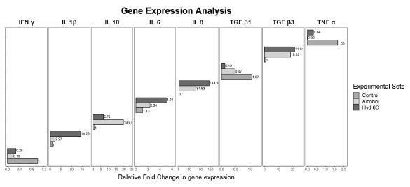
A panel of pro-and anti-inflammatory cytokines has been tested in this experiment (Figure 5) to observe their gene expression in response to the treatment of Hydrastis canadensis 6C dilution in alcohol against HepG2 cell line [18,19]. The decrease in relative fold change in gene expression of IFN ɣ(0.28), and pro-inflammatory cytokine TNF α(0.34) in comparison to the untreated cells (Control) justifies the anti-inflammatory effect of the drug dilution on the HepG2 cell line. On the other hand, the other pro-inflammatory cytokines, IL 6 (5.04), IL 8 (143.93), and IL 1β (14.26) support the null hypothesis, there is a minimal fold change increase in IL1β and IL 6, but in IL 8, there is a relative fold change increase of 143.93 in the drug dilution as compared to the control, which shows an opposite effect of the drug on these cytokine expression supporting the null hypothesis. However, important cytokines like IL-6 is pleotropic and as the outcome of the experiment was favourable, balancing action of IL-10 appears appropriate, as the relative fold change in gene expression has increased in IL 10 (6.76) and TGF β3 (21.61), which is also pleotropic, in comparison to the untreated cells, which supports the anti-inflammatory effect of the drug dilution on HepG2 cell line. The exact role of TGF β1 [20] requires elaborate studies, but in this study, the fold change observed in the drug dilution (0.12), decreased as compared to the control supporting the pro-inflammatory role of the cytokine, which in turn proves that the drug has an anti-inflammatory effect on the HepG2 cell line. Another observation from this study is that the fold change in alcohol-treated HepG2 cells is more than the drug-dilution-treated cells in the IL 10 (19.67) and TGF β1 (0.47) gene expression. This cytokine gene expression may be attributed to the direct hepatic cell damage by alcohol with reactive change to nullify it.
Table 1: Showing primers of different parameters studied in this experiment.
| Parameter |
Forward |
Backward |
| IFN-γ |
GAGTGTGGAGACCATCAAGGAAG |
TGCTTTGCGTTGGACATTCAAGTC |
| IL-6 |
AGACAGCCACTCACCTCTTCAG |
TTCTGCCAGTGCCTCTTTGCTG |
| IL-8 |
GAGAGTGATTGAGAGTGGACCAC |
CACAACCCTCTGCACCCAGTTT |
| IL-10 |
TCTCCGAGATGCCTTCAGCAGA |
TCAGACAAGGCTTGGCAACCCA |
| IL-1β |
CCACAGACCTTCCAGGAGAATG |
GTGCAGTTCAGTGATCGTACAGG |
| TGF-β1 |
TACCTGAACCCGTGTTGCTCTC |
GTTGCTGAGGTATCGCCAGGAA |
| TGF-β3 |
CTAAGCGGAATGAGCAGAGGATC |
TCTCAACAGCCACTCACGCACA |
| TNF-α |
CTCTTCTGCCTGCTGCACTTTG |
ATGGGCTACAGGCTTGTCACTC |
Table 2: PCR Cycle conditions in this experiment.
| Temperature |
Duration |
| Step 1: 95oC |
3.00 minutes |
| Step 2: 95oC |
15seconds |
| Step 3: 60oC |
30seconds |
| Step 2 and Step 3: 40 cycles |
Discussion
A study on HepG2 cells was performed to check the efficacy of the drug Hydrastis canadensis 6C on HepG2 liver cancer cells in comparison to control or untreated cells and alcohol-treated cells [21]. Cells challenged with Hydrastis canadensis 6C showed a prominent inhibition of cell proliferation when stained with methylene blue dye, and the cytotoxic nature of the drug was confirmed using the MTT assay, where the cell growth percentage (P<0.0001) significantly decreased, taking 95% confidence interval. Thus, these results indicated the anticancer activity of the drug by inhibiting cell proliferation. Apoptosis or programmed cell death plays an essential part in balancing cell death and cell proliferation and contributes to effective cancer therapy [22,23]. Many studies regarding anticancer activity have scientifically proven that apoptosis is the ultimate fate for anti-cancer therapy [24]. The apoptosis characteristic of the drug was further observed by the smear-like DNA band which is an outcome of DNA fragmentation and indicates apoptosis. Our results are suggesting that the cytotoxicity might be due to the apoptosis inductive effect indicative of anticancer activity of the drug Hydrastis canadensis 6C on the human Hepatoma (HepG2) cell line. We further studied the cytokine expression pattern, for observing the pro- or anti-inflammatory effect of the drug on the HepG2 cell line. The decrease in relative fold change in gene expression of pro-inflammatory cytokines, IFN ɣ, TNF α, and TGF β1 [21] indicates the anti-inflammatory effect of the drug while the relative fold change of other pro-inflammatory cytokines, IL-6, IL-8, and IL-1β increased, indicating a cytokine imbalance in the cancer cells. But the anti-inflammatory mediator’s expression increased, in comparison to the untreated cells, which supports the anti-inflammatory effect of the drug dilution on the HepG2 cell line. An increase in IL-10 expression was also seen which successfully counteracts the IL-6 expression, confirming the anti-inflammatory effect of the drug Hydrastis canadensis 6C on the HepG2 cell line. The efficacy of high-dilution bioactive chemicals never follows a linear relation between the concentration of the bioactive chemical and the effect of that chemical.
Again the succusion during preparation of this medicine may release silica nanoparticles from the glass surface, the bioactive chemical is probably entrapped inside the silica particles and released slowly afterwards. Thus at 6C ultradilution where an extremely scanty number of molecules are likely present, we are getting biological activities from that.
Conclusion
Our study design provides a background, confirming the cytotoxic, apoptotic, and anti-inflammatory nature of Hydrastis canadensis 6C – an alternative medicine on the HepG2 cell line. Thus, further studies are required to confirm the anticancer activity of the medicine on human hepatocellular carcinoma.
Declarations
Funding: There was no source of funding in this study.
Authors’ contribution: SD (Das), DC, PG, and KP designed the hypotheses and the experiments. SD (Dhar), DC, and BS performed the experiments and the analyses. SD (Dhar) has written the first draft of the manuscript. All authors participated in data interpretation and manuscript review and writing.
Competing interest: The authors declare no conflicts of interest.
References
- Arzumanian VA, et al. The Curious Case of the HepG2 Cell Line: 40 Years of Expertise. International journal of molecular sciences. 2021; 22(23): 13135.
- Chen S, et al. Mechanism study of goldenseal-associated DNA damage. Toxicology letters. 2013; 221(1): 64-72.
- Mandal SK. Goldenseal (Hydrastis canadensis L.) and its active constituents: A critical review of their efficacy and toxicological issues. Pharmacological research. 2020; 160: 105085.
- Chandrakar N, et al. Biological and physico-chemical characterization of ultra-high diluted Hydrastis canadensis and in vitro studies on mammalian cells. 2017; 10.1055/s-0043-104283.
- Wong RS. Apoptosis in cancer: From pathogenesis to treatment. Journal of experimental & clinical cancer research. 2011; 30(1): 87.
- Rimm EB, Stampfer MJ. Diet, lifestyle, and longevity--the next steps?. JAMA. 2004; 292(12): 1490-1492.
- Heber D, Bowerman S. Applying science to changing dietary patterns. The Journal of nutrition. 2001; 131(11 Suppl): 3078S-81S.
- Lu H, et al. Inflammation, a key event in cancer development. Molecular cancer research. 2006; 4(4): 221-233.
- Gutiérrez-Ruiz MC, et al. Cytokines, growth factors, and oxidative stress in HepG2 cells treated with ethanol, acetaldehyde, and LPS. Toxicology. 1999; 134(2-3): 197-207.
- Khuda-Bukhsh AR. Towards understanding molecular mechanisms of action of homeopathic drugs: An overview. Molecular and cellular biochemistry. 2003; 253(1-2): 339-345.
- Felice DL, et al. A modified methylene blue assay for accurate cell counting. J Funct Foods. 2009; 1: 109-118.
- Mosmann T. Rapid colorimetric assay for cellular growth and survival: Application to proliferation and cytotoxicity assays. Journal of immunological methods. 1983; 65(1-2): 55-63.
- Präbst K, et al. Basic Colorimetric Proliferation Assays: MTT, WST, and Resazurin. Methods in molecular biology. 2017; 1601: 1-17.
- Kamiloglu S, et al. Guidelines for Cell Viability Assays. Food Front. 2020; 1: 332-349.
- Rahbar Saadat Y, et al. An update to DNA ladder assay for apoptosis detection. BioImpacts, 2015; 5(1): 25-28.
- Khan SZ, et al. Heteroleptic Pd(II) Dithiocarbamates: Synthesis, Characterization, Packing and In Vitro Anticancer Activity against HeLa Cell Line. J. Coord. Chem. 2015; 68: 2539-2551.
- Al-Bakheit A, Abu-Qatouseh L. Sulforaphane from broccoli attenuates inflammatory hepcidin by reducing IL-6 secretion in human HepG2 cells. J. Funct. Foods. 2020; 75: 104210.
- Roy A, et al. Ultradiluted SARS-CoV-2 Spike Protein mitigates hyperinflammation in lung via ferritin and MMP-9 regulation in BALB/c mice. Virus Research. 2023; 329: 199091.
- Guerriero E, et al. Effects of lipoic acid, caffeic acid and a synthesized lipoyl-caffeic conjugate on human hepatoma cell lines. Molecules. 2011; 16(8): 6365-6377.
- Veldhoen M, Stockinger B. TGFbeta1, a Jack of all trades: the link with pro-inflammatory IL-17-producing T cells. Trends Immunol, 2006;27(8):358-61.
- Saha SK, et al. Ultra-highly diluted plant extracts of Hydrastis canadensis and Marsdenia condurango induce epigenetic modifications and alter gene expression profiles in HeLa cells in vitro. Journal of integrative medicine. 2015; 13(6): 400-411.
- Machana S, et al. Anticancer effect of the extracts from Polyalthia evecta against human hepatoma cell line (HepG2). Asian Pacific journal of tropical biomedicine. 2012; 2(5): 368-374.
- Gulecha V, Sivakuma T. Anticancer activity of Tephrosia purpurea and Ficus religiosa using MCF 7 cell lines. Asian Pacific journal of tropical medicine. 2011; 4(7): 526-529.
- De Bruin EC, Medema JP. Apoptosis and non-apoptotic deaths in cancer development and treatment response. Cancer treatment reviews. 2008; 34(8): 737-749.