Introduction
Globally, Breast Cancer (BC) is one of the most common disease associated with women heath due to poor early prognosis and diagnosis. Genetic and epigenetic factors make the etiopathology of BC more complex due to variable expression of Epithelial - Mesenchymal Transition (EMT) markers-Sox4, EpCAM and CK19 in Circulating Tumor Cells (CTCs) isolated from variety of tumors during progression of disease [1-4]. Sox4 gene belong to Sry-related High Mobility-Group (HMG) domain and act as early transcription factor to regulate cellular - differentiation process and to decide the fate of tumours during metastasis [5]. Sox4 gene assigned on chromosome- 6p22.3 with single exon that encodes 47KD protein comprising of 474 amino acids residues regulating growing progenitor cells through multiple signalling, among Transforming Growth Factors (TGF) or Protein Kinase (PKC) are responsible for the activation of oncogene (KRAS). Sox4 induces aggressive behaviour after prolonged exposure during transition of epithelial cells by TGFβ signalling in differentiating tumor cells [6,7].
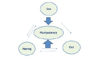
During tumorigenesis, Sox4 may also act as potential biomarkers for early diagnosis and their gene-expression varying with different age and ethnic groups depending upon invasion to negihbouring tissues (organs) and migration of CTCs [8,9]. Nanog, an another transcription factor, and is essential to maintain pluripotency with variable expression suppresses the differentiation of embryonic stem cells due to self-renewal property during angiogenesis [10]. Hence, the curiosity has been developed with the aim to explore the functional etiopathology of Sox4 maintaining pluripotency either alone or in synergistic fashion either with Oct4 or Nanog and also evaluate the frequency of DNACNVs in individual cancer patients to explore the genetic susceptibility. The present study is small but have great potential to the clinicians for early prognosis, diagnosis and followed by timely management using stem cells targeted therapeutics as a source of regenerative medicine.
Materials and Methods
Present study includes clinically diagnosed cases (n=38) of breast cancer patients and controls (n=38) were used for the present study. The study is approved by Institute Ethical Committee (IEC), All India Institute of Medical Sciences Patna (Bihar). Blood samples (1.0 ml) were collected in a EDTA vial after consent form the patients. Genomic DNA was isolated using kit protocol and half of the sample (cells) were fixed in RNAzole for isolation of RNA for quantification by nanodrop spectrophotometer. The samples were stored at -20⁰C, till further analysis of stem cells markers using PCR technique with specific forward and reverse primers on agarose gel (1.5%) electrophoresis.
Characterization of Sox4, Oct4 and Nanog.
Table-1 showing the PCR based strategy for Sox4, Oct4 and Nanog analysis along with their annealing temperature using specific primers. The nucleotide sequences of the primers further confirm by NCBI-BLAST (http://blast.ncbi.nlm.nih.gov.). Total volume (25 μl) of PCR was achieved in reaction mixture containing 5X Green GoTaq PCR reaction buffer, dNTPs (10 mM), 1 μl each of 10 pmol of Oct4, Nanog, Sox2 and Sox4 primer (forward & reverse), 0.2 μl of GoTaq DNA polymerase (5 U/μl) and 50 ng DNA in thermocycler (Agilent Technologies, Sure Cycler 8800). Reaction summary was carried out in 35 cycles comprising, hot start at 95°C for 5 minutes, denaturation at 95oC for 30 seconds, annealing at 57.2oC for 30 seconds, elongation at 72oC for 30 seconds, followed by final elongation at 72°C for 8 minutes. The amplified PCR product of the candidate genes were characterized individually on agarose gel electrophoresis after ethidium bromide staining. The individual band intensity was measured by densitometry to evaluates the frequency of DNACNVs using Image Lab inbuilt Software of Gel Doc system.
Table 1: PCR strategy was used for the characterization of stem cell marker in breast cancer patients.
| Stem cell Marker |
Forward & Reverse Primer Sequences (5’ → 3’) |
Annealing Temp (oC) |
References |
| Sox4 |
f5’-GGTCTCTAGTTCTTGCACGCTC-3' |
57.2 |
Jafarnejad, et al (9) |
| Sox4 |
r5’-CGGAATCGGCACTAAGGAG-3' |
| Oct4 |
f5-‘GACCATCTGCCGCTTTGAG-3' |
60.0 |
Henderson, et al (11) |
| Oct4 |
r5;-CCCCCTGCCCCCATTCCTA-3' |
| Nanog3 |
f5'-CTGTGATTTGTGGGCCTG AA-3' |
56.0 |
Nettersheim, et al (10) |
| Nanog3 |
r5'-TGTTTGCCTTTGGGACTGGT-3' |
Table 2: Statistical analysis showing the frequency (%), O.R and C.I for Oct4, Nanog and Sox4 in breast cancer patients with controls.
| Type of genes |
Mean ± SD |
Gene expression |
Frequency (%) cases controls |
C.I. at 95% max min |
OR |
p-value |
| Sox 4 |
2,127.3 ± 368.6 |
Over-expression |
6 (75.0) |
1 (14.3) |
5.11 |
0.53 |
18.0 |
0.018* |
| 1618.9 ± 216.0 |
Under-expression |
2 (25.0) |
6 (85.7) |
11.42 |
0.15 |
| Oct-4 |
2099.0 ± 106.3 |
Over-expression |
6 (75.0) |
4 (57.1) |
33.62 |
2.68 |
2.25 |
0.88 |
| 2108.5 ± 134.5 |
Under-expression |
2 (25.0) |
3 (42.9) |
7.7 |
1.59 |
| Nanog |
2191.3 ± 223.1 |
Over expression |
4 (50.0) |
4 (57.1) |
8.46 |
0.529 |
0.75 |
0.001* |
| 1678.5 ± 265.9 |
Under-expression |
4 (50.0) |
3 (42.9) |
4.93 |
0.474 |
DNA Copy Number Variations in Sox4, Oct4 and Nanog
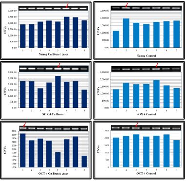
Fluorescence stain, ethidium bromide was used for the visualization of individual bands intensity and densitometry analysis were carried for three stem cell markers- Sox4, Oct and Nanog in BC patients and compare with age match controls using Image lab software 5.1 of inbuilt Gel Doc system (Bio Rad USA). Data was evaluated in the terms of either over-expression (up regulation) or under-expression (down regulation) and bar diagram was constructed for visualization of the frequency distribution of DNACNVs of stem cell markers to determine the genetic susceptibility during advancement of disease in cancer patients.
Statistical analysis
Chi-square test (two tailed) was used for evaluation significant (p-value) difference between cases and controls. Data was also calculated mean ± standard deviation, standard error, Odd-Ratio (O.R) and Confidence Intervals (C.I) at 95% (max & min) values to find the interaction between two variables.
Results
Table-2 showing the detail analysis of the data of three pluripotent stem cell markers - Sox4, Oct4 and Nanog along with the frequency of DNACNVs in BC patients. Interestingly, Sox4 showing significant over-expression with respect to controls as shown in Figure 1C & 1D. The calculated values of mean±SD (2127.3±36.8), confidence interval C.I) varies between 511-0.53 with odd ratio 18.0 (Table 2). Similarly, bar diagram of DNACNVs showing over-expression in Oct4 and Nanog with calculated mean±SD values 2099.0±106.3, 2191.3±223.1, confidence interval varying between 33.62-2.68, 8.46-0.5 with O.R 2.25, 0.75, respectively as depicted in Figure 1A,B,E&F(arrow). Interestingly, Nanog showing again highly significant (p<0.001) differences (over-expression) with calculated value of O.R is 0.75 and in C.I at 95% intervals that varying between 8.46 to 0.529 with respect to controls.
Discussion
Stem cell biology is the frontier area of regenerative medicine and are known to participate in cellular-differentiation, invasion and migration in variety of diseases other than tumorigenesis [12]. Etiopathology of stem cells (progenitor cell) with functional aspects has been poorly documented in literature and antitumor agent or environmental mutagen (arsenic) increase risk of developing cancer after genetic and epigenetic modification [13]. Present study is interesting and significant finding in DNA copy number of Sox4 confirm to induce metastasis during tumorigenesis. In earlier study of the same group emphasize that stem cells like Oct4, Nanog and Sox2 play a relevant role in normal differentiation of germ-cell and their expression maintain “central- dogma” of pluripotency during organogenesis other than cancer [14,15]. In fact, Sox proteins act as architect to decide the fate of early developmental processes including sex-determination, neural , chondrogenesis and cardic development [16,17]. Zhang et al. also suggested that Sox4 expression is also influence by chemokine transmembrane receptor [8]. The most interesting part of the present study showing significant relationship of Sox4 between DNACNVs and over-expression that confirming the growth, (proliferation), migration and invasion cells to induce metastasis BC patients.
Sox-4, an early transcription factor and is regulatory molecule during developmental process through binding to the adjacent site of the DNA with other partner for activation or repression, that confirm association with Nanog, might be the “good” partner for maintain pluripotency in BC patients [12]. The ectopic expression of Sox4 increase the genetic susceptibility after using Paclitaxel (PTX) or Chemokine Transmembrane Recptor-7 (CXCR7) pathway to decrease metastasis [8]. During Transition of Epithelial to Mesenchymal cells (EMT) involves another regulatory pathway that includes CK19, EpCAM and Sox4, and modulates gene-expression through TGF β in synchronize manner, suggesting induce tissues specific genetic susceptibility during progression of disease [1,18]. Over-expression of Sox4 is resposible for aggressive behivor during transition of epithelial to mesenchymal cells through EpCAM or CK19 in liver and pancreatic tumor [19-20]. Sox4 mRNA expression is dependent on TGF-β, a pleutrophic cytokine has been associated with glioma and pituatry cells through Sox2, suggesting multilineage differentiation quality of stem cells [21,22]. Data of the present study confirm that functional activity of Sox4 is not alone but responsible with synergistic fashion with Nanog together inhibit metastasis in during progression of disease. Although, over-expression of Nanog as transcription factor becomes relevant for differentiation of tumor cells, but exact mechanism how to accelerate mammary tumorigenesis is still lacking [23]. Present study of DNACNVs also show over-expression of Oct4, but showing lacking significance difference with respect to controls, suggesting fail to form complex either with Sox4 or Nanog resulting catastrophe regulation in maintaining pluripotency. The mimicry (up/down regulation) of stem cell markers further confirmed by DNACNVs in individual patients occurs either due to different stages of malignancy or varying genetic susceptibility. Another aspect of the variation of DANCNVs is genetic heterogenicity, might be another aspect for unequal proliferation of circulating tumor cells during migration, invasion to the neighbouring tissues fail to maintain pluripotency in BC patients.
Conclusion
Tissue culture engineering based on stem cell tumour biology has great value for the clinicians in therapeutics as a source of regenerative medicine in variety of disease like chronic wound healing and early management of cancer. In order to develop effective treatment approach, there is an insistent need to comprehend the causes of heterogeneity in cancer patients with its cellular and molecular traits are as follow - 1) the functional activity of Sox4 confirm that act as synergistic fashion with Nanog, 2) significant variation of DNACNVs increases genetic susceptibility due to different stages of cancer either pre or post metastatic, and 3) The aggressive behaviour of epithelial to mesenchymal cell confirm by DNACNVs of Sox4 concluding that stem cell therapy act as source of regenerative medicine may help for cancer patients as supportive tool without any side effects.
Declarations
Acknowledgement: Author (AKS) is thankful to the Department of Science and Technology, New Delhi for providing Research Grant. Moreover, we are also thankfully acknowledge to the patients who participated in this project.
Funding: This study was funded by Ministry of Science and Technology, New Delhi, India (Grant number: DST/TDP/BTDT16/2021)
Availability of data and materials: The data presented in this study are available on request from the corresponding author.
Authors’ contributions: AKS executed experimental design, implementation and proof reading.
Ethics approval and consent to participate: Study was approved by Institute ethical committee of AIIMS Patna (AIIMS/Pat/IRC/2020/610), and informed consent form was dually signed by patients.
Patient consent for publication: Patients consent form was dually signed by patients involved in study.
Competing interests: There are no competing interests among the authors.
References
- Saxena Ajit K, Tiwari M, Singh, P, Singh V. Comprehensive Mutational Spectra of Epithelial Mesenchymal Transition Markers-SOX4, EpCAM, CK19 and their Impact of Methylene Tetrahydrofolate Reductase C677T Gene Polymorphism Associated Risk Factor Modified by BRCA and TGFβR1 Genes in pre/post -menopausal Cases of Breast International Journal of Cancer. Medicine. 2024; 7(1): 158-182.
- Cohen SJ, et al. Relationship of Circulating Tumor Cells to Tumor Re sponse , Progression-Free Survival, and Overall Survival in Patients with Metastatic Colorectal Cancer. Journal of Clinical Oncology. 2008; 26: 3213-3221.
- De Bono JS, et al. Circulating Tumor Cells Predict Survival Benefit from Treatment in Metastatic Castration-Resistant Prostate Cancer. Clinical Cancer Research. 2008; 14: 6302-6309.
- Cristofanilli M, et al. Circulating Tumor Cells, Disease Progression, and Survival in Metastatic Breast Cancer. The New England Journal of Medicine. 2004; 351: 781-791.
- Perou CM, Sørlie T, Eisen MB, van de Rijn M, Jeffrey SS, Rees, et al. Molecular portraits of human breast tumours. Nature. 2000; 406(6797): 747-752.
- Ikushima H, Miyazono K. TGFβ signalling a complex web in cancer progression. Nature Rev. Cancer 2010; 10: 415-426.
- Lefebvre V, Dumitriu B, Penzo-Mendez A, Han Y, Pallavi B. Control of cell fate and differentiation by Sry-related high mobility-group box (Sox) transcription factors. Int J Biochem Cell Biol. 2007; 39: 2195-2214.
- Zhang J, Xiao C, Feng Z, Gong Y, Sun B, et al. SOX4 promotes the growth and metastasis of breast cancer. Cancer Cell International. 2020; 20(1): 1-11.
- Jafarnejad SM, Wani AA, Martinka M, Li G. Prognostic significance of Sox4 expression in human cutaneous melanoma and its role in cell migration and invasion. American journal of pathology. 2010; 177(6): 2741-2752.
- Nettersheim D, Biermann K, Gillis AJM, Steger K, Looijenga LHJ, et al NANOG promoter methylation and expression correlation during normal and malignant human germ cell development. Epigenetics. 2011; 6(1): 114-122.
- Henderson JK, Draper JS, Baillie HS, Fishel S, Thomson JA, et al. Preimplantation Human Embryos and Embryonic Stem Cells Show Comparable Expression of Stage‐Specific Embryonic Antigens. STEM CELLS. 2002; 20(4): 329-337.
- Prior HM, Walter MA. SOX Genes: Architects of Development. Molecular Medicine. 1996; 2(4): 405-412.
- Lu X, Mazur SJ, Lin T, Appella E, Xu Y. The pluripotency factor Nanog promotes breast cancer tumorigenesis and metastasis. Oncogene. 2014; 33(20): 2655-64. doi: 10.1038/onc.2013.209. Epub 2013 Jun 17. PMID: 23770853; PMCID: PMC3925756.Saxena.
- Ajit K, Pandey S, Pandey LK. Genetic diversity of stem cells and their functional impact on the development of neural tube defects in population of Eastern India. Genetics and Molecular Research. 2013; 12(3): 2380-2380.
- Mehta GA, Parker JS, Silva GO, Hoadley KA, Perou CM, et al. Amplification of SOX4 promotes PI3K/Akt signalling in human breast cancer. Breast cancer research and treatment. 2017; 162(3): 439-450. https://doi.org/10.1007/s10549-017-4139-2.
- Kamachi Y, Kondoh H. Sox proteins: Regulators of cell fate specification and differentiation. Development. 2013; 140(20): 4129-4144.
- Saxena Ajit Kumar, Shalini, K Manoj. Flowcytometric based Analysis of Sox4 Gene Expression-An Early Transcription Factor Influenced by 5-Azacytidine and Compare with EpCAM in Circulating Tumor Cells Isolated from Breast Cancer Patients. Journal of Oncology Research and Therapy. 2024; 9(03): 1-7.
- Vervoort SJ, De Jong OG, Roukens MG, Frederiks CL, Vermeulen JF, et al. Global transcriptional analysis identifies a novel role for SOX4 in tumor-induced angiogenesis. eLife. 2018; 7: e27706. https://doi.org/10.7554/eLife.27706.
- Zhang J, Liang Q, Lei Y, Yao M, Li L, et al. SOX4 Induces Epithelial - Mesenchymal Transition and Contributes to Breast Cancer Progression. 2012; 4597-4609. https://doi.org/10.1158/0008-5472.CAN-12-1045.
- Thein DC, Thalhammer JM, Hartwig AC, Crenshaw EB, Lefebvre V, et al. The closely related transcription factors Sox4 and Sox11 function as survival factors during spinal cord development. Journal of Neurochemistry. 2010; 115(1): 131-141. https://doi.org/10.1111/j.1471-4159.2010.06910.x.
- Song GD, Sun Y, Shen H, Li W. SOX4 overexpression is a novel biomarker of malignant status and poor prognosis in breast cancer patients. Tumor Biology. 2015; 36(6): 4167-4173. https://doi.org/10.1007/s13277-015-3051-9.
- Ikushima H, Todo T, Ino Y, Takahashi M, Miyazawa K, et al. Autocrine TGF-beta signaling maintains tumorigenicity of glioma-initiating cells through Sry-related HMG-box factors. Cell Stem Cell. 2009; 5(5): 504-14. doi: 10.1016/j.stem.2009.08.018. Erratum in: Cell Stem Cell. 2009; 5(6): 666.
- Kuwahara M, Yamashita M, Shinoda K, Tofukuji S, Onodera A, et al. The transcription factor Sox4 is a downstream target of signalling by the cytokine TGF-β and suppresses T(H)2 differentiation. Nat Immunology. 2012; 13(8): 778-86. doi: 10.1038/ni.2362. PMID: 22751141
- Lee S, Osmanbeyoglu HU. Chromatin accessibility landscape and active transcription factors in primary human invasive lobular and ductal breast carcinomas. Breast Cancer Res. 2022; 24(1): 54. doi: 10.1186/s13058-022-01550-y.